Geochemistry and Mineralogy Core Facility
This research core offers instrumentation including SEM/EDS, XRD, polarized-light microscopy, and a fluid-inclusion geothermometry stage for the elemental and mineralogical characterization of solids.
Imaging, mineralogical and elemental analyses

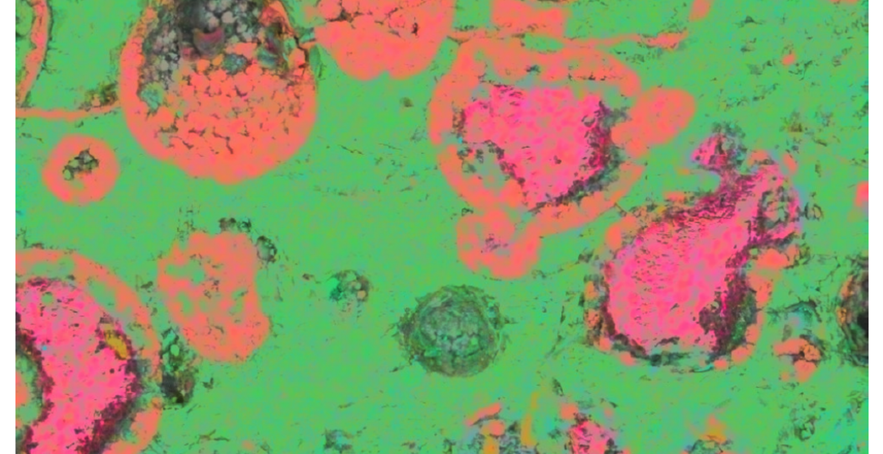
Cocolith microfossils from the Gulf of California.
This research core offers instrumentation including SEM/EDS, XRD, polarized-light microscopy, and a fluid-inclusion geothermometry stage for the elemental and mineralogical characterization of solids.
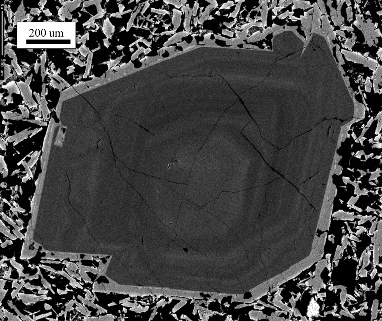
The Scanning Electron Microscopy (SEM) is equipped with the energy dispersive X-Ray spectroscopy (EDS). The SEM/EDS system is used for high-resolution imaging and elemental analysis of a variety of solid sample types, including rocks and minerals, concrete and mortars, electronic and semiconductor materials, metals and alloys, synthetic catalysts, pharmaceuticals, and dried biological specimens.
The X-ray diffractometer (XRD) is used for routine identification of powdered samples of crystalline materials, singly or in mixtures, such as minerals in rocks, ceramics, concrete and mortar phases, and synthetic crystalline materials. MDI Jade software is available for processing the XRD data.
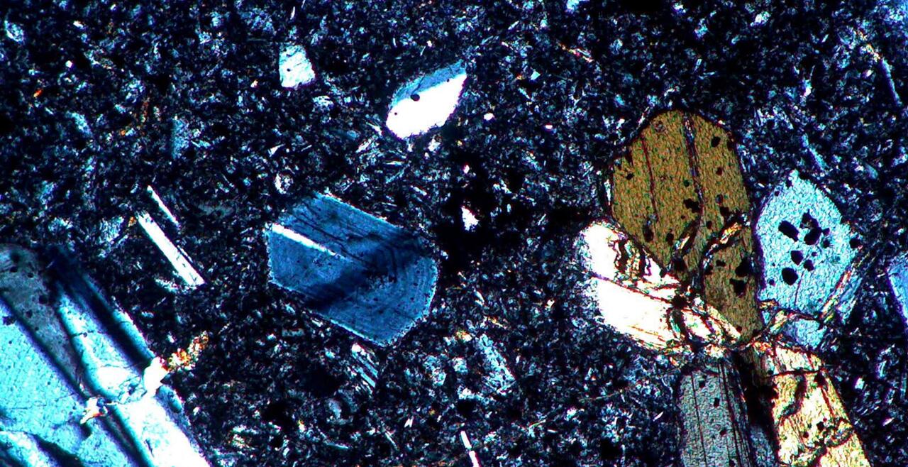
The lab also houses a Nikon Optiphot-pol research petrographic microscope equipped for transmitted and reflected light microscopy, digital photomicrography, and determination of optical properties of crystals using a spindle stage and Excalibr software.
A Fluid Inc. gas-flow fluid-inclusion geothermometry stage is also available. This instrument can heat or cool fluid inclusions in a transparent mineral to determine the minimum temperature at which the fluid was trapped during crystal growth (by heating until the vapor bubble disappears), and to determine the salinity of the fluid (by freezing point depression). Being mounted on a polarized light microscope, it can also be used to investigate temperature-dependent reactions, melting, and crystallization processes.
Petrographic microscopes with ProMicrom 3.0 digital camera and image mosaic software is available to create gigapixel imagery from material samples.
Fee Structure
There are UMKC, external academic and external commercial rates for equipment use and sample preparation and processing. Additional charges for consumables and color printing may apply. Digital imagery is included.
Imagery from UMKC SEM and Petrographic Mosaic system
Recent research images